Imaging
RXDC X-RAY UNIT
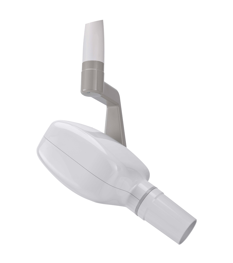
RXDC, the user-friendly intra-oral x-ray unit
Technology at the service of innovation.
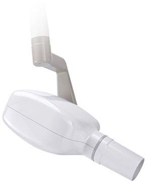
The RXDC provides top-drawer imaging with outstanding detail thanks to a constant potential high frequency generator (DC). With a very small focal spot (0.4 mm) it’s possible to obtain sharp images with ultra-high definition.
The RXDC maximises both imaging performance and patient comfort while significantly lowering the X-ray doses he/she is exposed to. Highly versatile and simple to install, the X-ray unit has arms with an integrated self-balancing system that allows them to be pointed in 4 directions - available in the following lengths: 40, 60 and 90 cm. Thanks also to the protractor with graduated scale, positioning of arms and tube head is stable and easy.
X-VS SENSOR
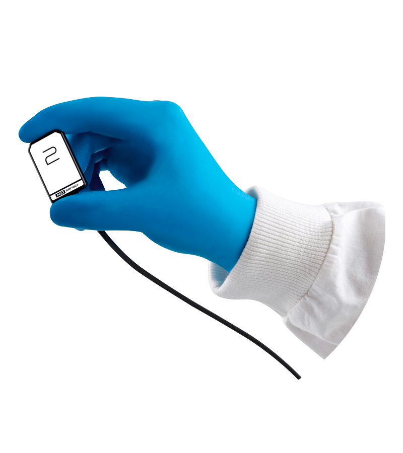
X-VS, Intra-oral sensor for direct USP high definition images
Reduced dimensions and a maximum active area ensure advanced-ray diagnosis, The X-VS is equipped with a sensor, available in two sizes -ergonomic and with smoothed edges - which adapt it to the anatomy of the patient’s oral cavity, guaranteeing outstanding positioning comfort.
Advanced filters and Multi Level vision
The Multi-Layer-Filters function lets dentists simultaneously capture, display and share a set of images (up to 5), each with a specific improvement that can be used to highlight various anatomical details with different degrees of sharpness. After image capture, users can customise image contrast to suit their diagnostic or visual preferences, allowing for improved diagnosis.
Software iRYS
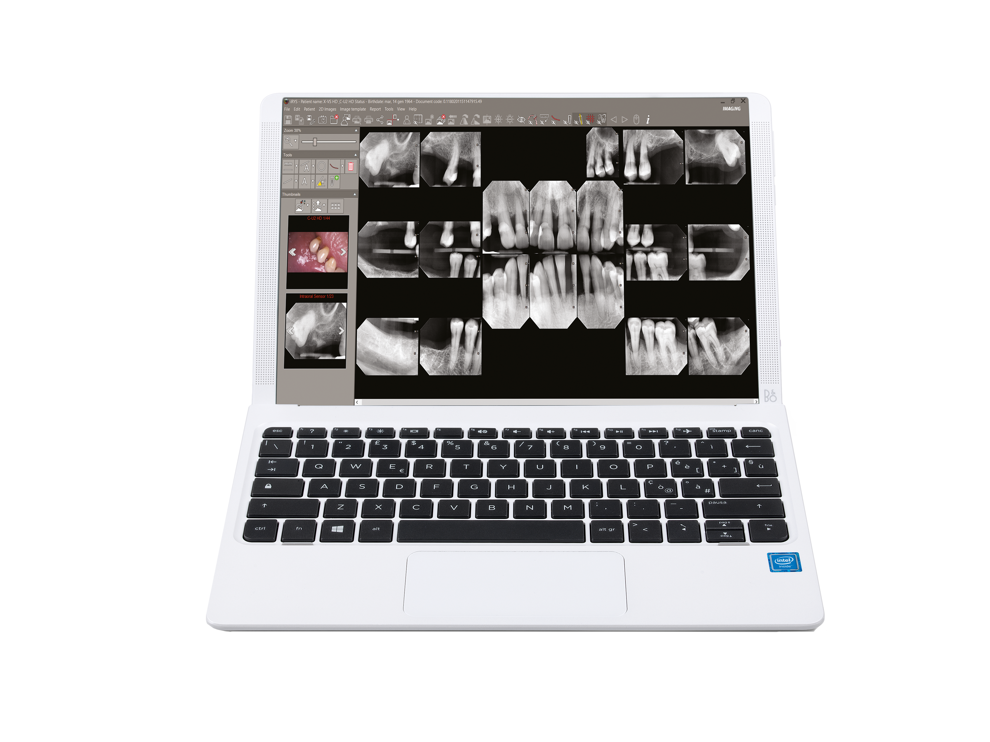
iRYS, the all-in-one software platform for 2D and 3D imaging, is DATA PROTECTION certified and IHE compliant with DICOM networks.
iRYS is a tool that provides dentists with an array of functions that lets them view, process and share captured images - directly from the dedicated workstation - with the computers in the dental practice and the iRYS Viewer app for iPADs.
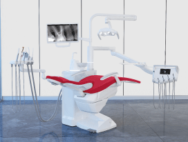
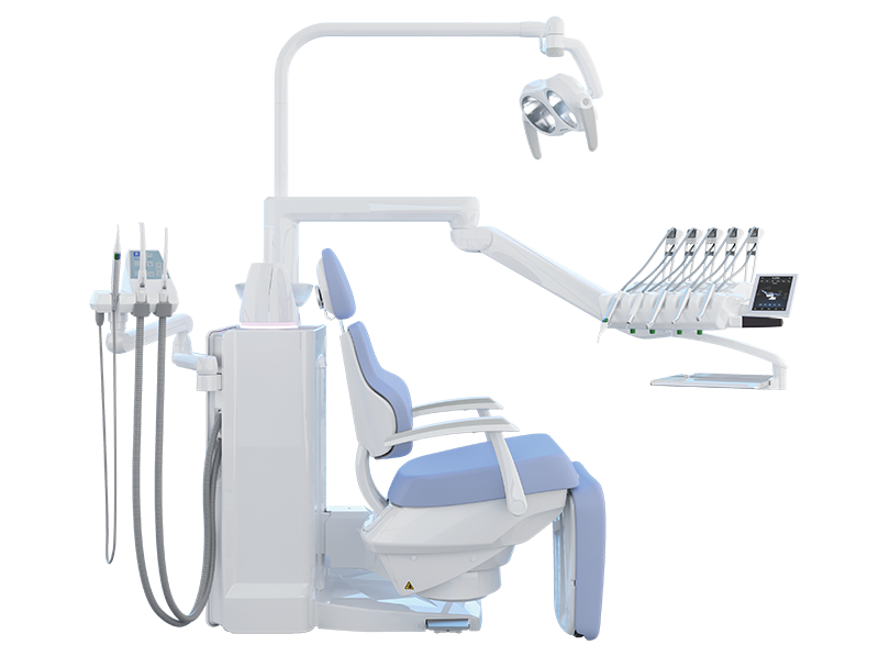
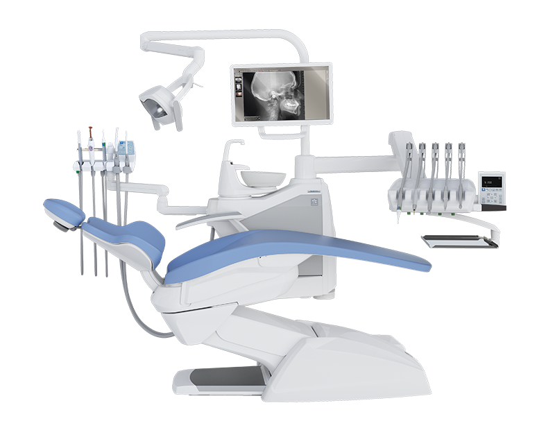
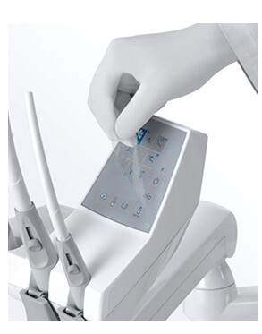
iRYS lets dentists manage diagnostic images in the patients’ medical records. The latter are shared on the network and can also be accessed from the dental unit, which is equipped with a multimedia card and a FullTouch Multimedia control panel via which new images captured by the unit itself can be saved.
Functional features:
Image management in the patient’s medical records
2D/3D Multi-desktop with advanced, customizable filters
Dental status template with session scenario save function
Image is associated with the anatomical region of the dentition X-ray dose log
Compatibility with third-party software and devices
Processed images can be shared via the viewer




.svg)
.svg)
